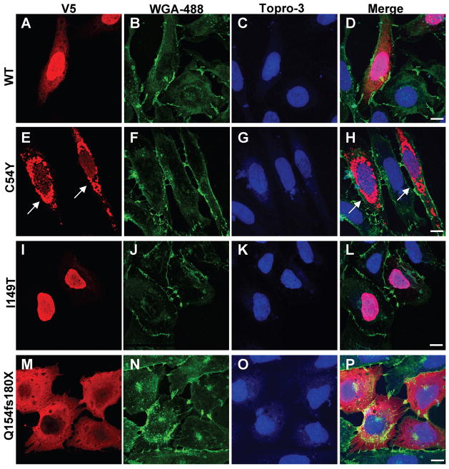
Fig. 3. Nuclear localization is altered in some DYT6 mutants.
V5-tagged THAP1 variants in U2OS cells localized via immunofluorescence using anti-V5 monoclonal antibody (red), wheatgerm agglutinin-Alexa488 (membranes; green), TO-PRO®-3-iodide (nuclei; blue). (A–D) V5-wtTHAP1 was expressed in both nucleus and cytosol of transfected cells. (E–H) The C54Y mutant was frequently observed in robustly labeled, perinuclear inclusions (arrows) which were never observed in cells expressing wtTHAP1 or any of the other DYT6 mutants. (I–L) Although the I149T mutation falls within the predicted NLS, it did not disrupt nuclear import as shown by strong nuclear labeling in cells expressing this mutant. (M–P) The Q154fs180X mutant was localized exclusively within the cytoplasm, indicating impaired nuclear entry due to disruption of the NLS. The remaining mutants (F81L, ΔF132, T142A, A166T) produced staining patterns similar to that of wtTHAP1 (Fig. S1). Images shown were captured by laser confocal microscopy at 100X final magnification under oil immersion. Scale bars = 10 μM.

