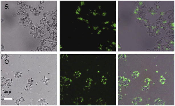
Figure 8.
Fluorescent images of live human ovarian carcinoma cells (OVCAR3, top row) and human colonic adenocarcinoma cells (HT29, bottom row) after incubation with folic acid modified PEI/NaYF4 UCNPs. The left rows are images in bright field, the middle rows are fluorescent images in dark field, and the right rows are overlays of the left and middle rows111

