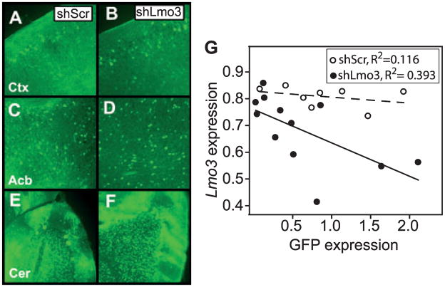
Fig. 3.
Characterization of transgenic mice expressing shLmo3.8 or shScr. (A–F) Green fluorescent protein (GFP) fluorescence in representative sagittal adult brain sections of transgenic mice infected at the single-cell embryo stage with lentivirus encoding shLmo3.8 (B, D, F) or shScr (A, C, E). GFP expression from the viral vector is visible in cell bodies (bright spots) and processes of neurons throughout the brain, including the cortex (A, B, Ctx), nucleus accumbens (C, D, Acb), and cerebellum (E, F, Cer). Hazy green areas represent background autofluorescence from the tissue, rather than infection per se. (G) Expression of GFP and Lmo3 transcript levels were negatively correlated in the forebrains of transgenic shLmo3.8 (closed circles, n = 12), but not shScr (open circles, n = 8) mice. GFP (x-axis) and Lmo3 (y-axis) expression values were normalized relative to expression of the housekeeping gene Gapdh.

