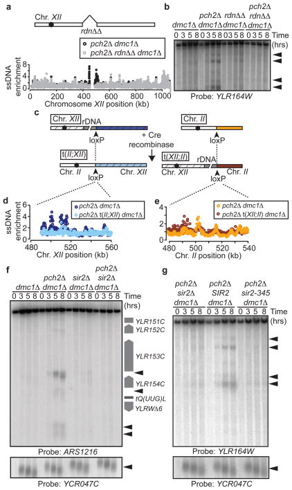
Figure 4. rDNA chromatin promotes DSB formation.
a, Schematic of rdnΔΔ strain and ssDNA profiles of region flanking the right rDNA border in pch2Δ dmc1Δ (H2629, black) and pch2Δ rdnΔΔ dmc1Δ (H4737, grey) cells. b, Southern blot analysis of the right rDNA flank in dmc1Δ (H118), pch2Δ dmc1Δ (H2629), rdnΔΔ dmc1Δ (H4736), and pch2Δ rdnΔΔ dmc1Δ (H4737) cells. c, Strategy used to generate chromosomal translocations between chromosomes XII and II. d and e, ssDNA profiles of strains containing the XII;II translocation. In d, the depicted region is next to the rDNA in pch2Δ dmc1Δ (H2629) cells (dark blue) and next to the left arm of chromosome II in pch2Δ t(II;XII) dmc1Δ (H4798) cells (light blue). In e, the depicted region is located on chromosome II in pch2Δ dmc1Δ cells (orange) and next to rDNA in pch2Δ t(XII;II) dmc1Δ cells (dark red). f, Southern blot of left rDNA flank (HindIII; probe: ARS1216) and YCR047C in dmc1Δ (H118), pch2Δ dmc1Δ (H2629), sir2Δ dmc1Δ (H2953) and pch2Δ sir2Δ dmc1Δ (H3038) cells. g, Southern blot of the right rDNA flank and YCR047C in pch2Δ sir2Δ dmc1Δ (H3262), pch2Δ sir2Δ dmc1Δ leu2::SIR2 (H3261), and pch2Δ sir2Δ dmc1Δ leu2::sir2-345 (H3282) cells.

