Figure 2.
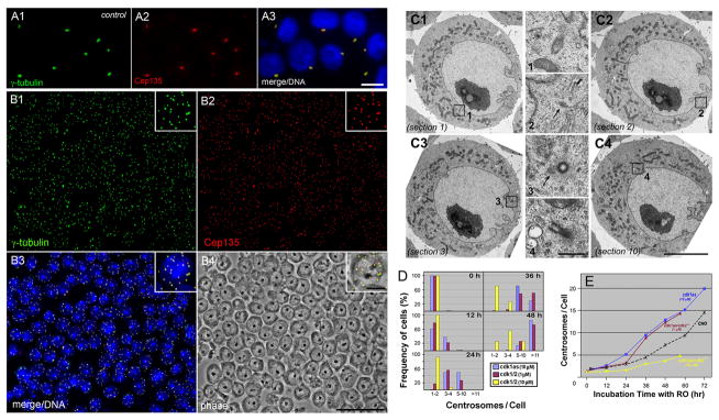
Formation of multiple centrosomes in DT40 cells. A, B: Immunostaining of cdk1as cells with anti-γ-tubulin (A1, B1) and anti-Cep135 (A2, B2) antibodies. Cells were treated with (B) and without (A) 10 μM PP1 for 48 hr. Bars, 10 μm (A and inlets of B) and 50 μm (B). C: Electron micrographs of PP1-treated cdk1as cells containing multiple centrioles. Four out of ten serial thin sections are shown in C1 to C4. 4–5 centrioles/centriole-like structures are detected in total ~1 μm thickness of sections as outlined in C1 to C4. Each structure is seen at a higher magnification in middle columns numbered 1 to 4. Note that centrioles at the juxanuclear reposition are spread widely apart from each other. Arrows indicate a position of centriole-like structures. Bars, 5 μm (C1 to C4) and 0.5 μm (1 to 4). D: Frequency histograms of cdk1as (blue) and cdk1/2 (cdk1as/cdk2−/−; brown and yellow) cells containing 1–2, 3–4, 5–10, and over 11 centrosomes after treatment with 1 or 10 μM PP1 for different periods of time. E: Time course analysis of centrosome amplification in CHO and DT40 cells. After incubation of CHO cells with 10 μM RO (black dots), cdk1as cells with 10 μM PP1 (blue), and cdk1as/cdk2−/− cells with either 1 μM (brown) or 10 μM (yellow) PP1 for various times, cells were fixed and double stained with γ-tubulin and Cep135 antibodies to count the number of centrosomes.

