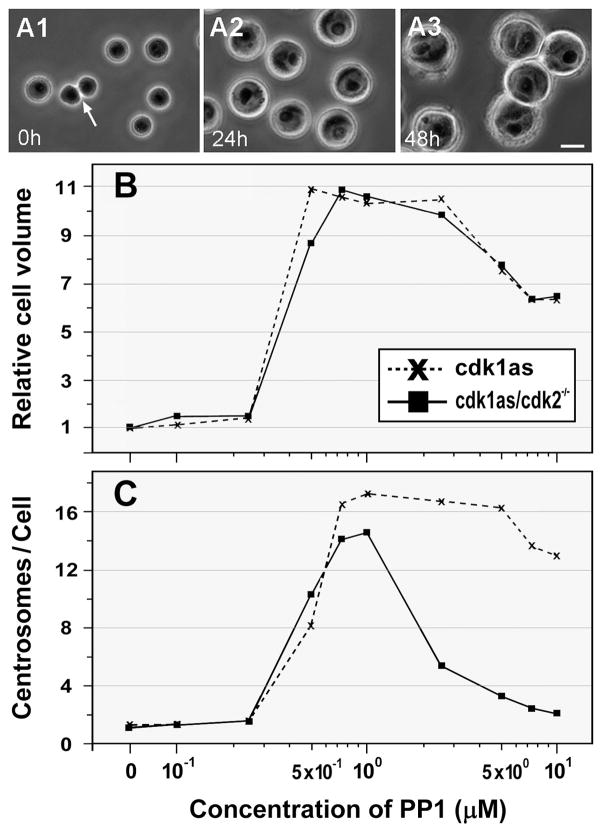
Figure 5.
Analysis of cell size and numbers of centrosomes in DT40 cells with inactivated Cdks. A: Phase-contrast microscopy of cdk1as cells treated with 10 μM PP1 for 0 (A1), 24 (A2), and 48 (A3) hr. An arrow indicates two daughter cells formed after cell division. Bar, 10 μm. B: Relative cell volume of cdk1as and cdk1as/cdk2−/− cells after incubation with various concentrations of PP1 for 48 hr. C: The average number of centrosomes induced in cdk1as and cdk1as/cdk2−/− cells after treatment for 48 hr with various concentrations of PP1. Centrosome overduplication is blocked in cdk1as/cdk2−/− cells treated with over 1 μM PP1.

