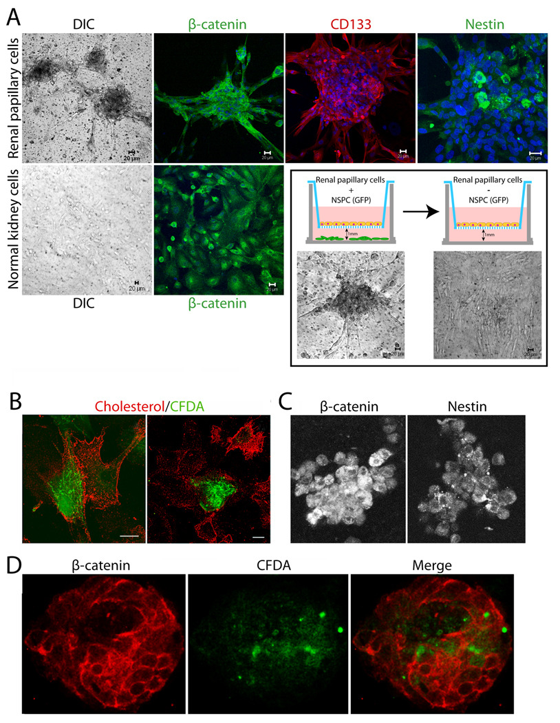
Figure 5. Human papillary cells associate with cortical tubular epithelia and undergo significant morphogenesis in co-culture systems.
(A) CD133/1+ papillary cells were grown in the presence or absence of GFP-neural stem/progenitor cells for 3–4 d, in defined serum-free neural stem cell medium as illustrated in the cartoon. Top row left to right shows human papillary cells cultured on filters above GFP-neural progenitors: DIC image of spheroids, β-catenin (green, single slice), CD133 (red, 3D reconstruction of complete Z-stack) and nestin (green, single slice) stained spheroids. Bottom row left two panels show primary normal human cortical tubular epithelia cultured on filters above GFP-neural progenitors: DIC image, β-catenin (green). Nuclei stained with Hoechst are in blue. DIC images within the outlined box (below cartoons) show flattening of papillary spheroids when filter cultures are transferred to wells lacking neural stem cells for 24 h. (B–D) Papillary cells loaded with CFDA fluorescent dye (green) readily form contacts with primary normal human cortical tubular cells in co-culture experiments on plastic dishes. Staining for cholesterol using perfringolysin O (red) was used to identify all cells in the culture. (B) Primary tubular epithelia (red) and papillary cells (green and red) interact closely on plastic dishes irrespective of whether tubular epithelia are from the same or a different patient origin as papillary cells. (C) Papillary cells grown 10 d in collagen gel cultures stained for nestin and β-catenin. (D) Primary tubular epithelia (red) and CD133+ papillary cells (green and red) grown in collagen gel co-cultures interact and form tubulospherical structures with hollow cores. See supplemental movie 1 to view entire Z-stack.

