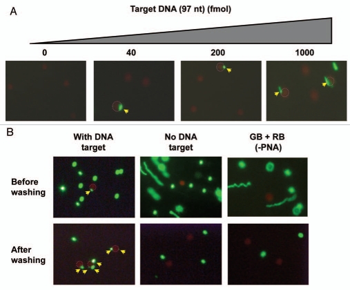
Figure 2.
Detection of a 97 nt target DNA with a pair of PNA-Beads. Two PNA beads (red beads (RB) with PNA3534 and green beads (GB) with PNA3533), prepared as illustrated in Figure 1, were incubated with the target DNA and subsequently analyzed by fluorescence microscopy. RBs, showing a co-localization with GB(s) are circled with dotted red line, and the co-localized GBs are indicated by yellow arrows. (A) Dose dependent detection of target DNA (0–1,000 fmol). (B) The complex formation was tested in the presence or absence of target DNA (40 fmol) with or without PNA probe. The samples were subjected to fluorescence microscopy before and after magnetic enrichment. A two step hybridization was performed to assure Red beads/DNA complexation first as the Red beads have less DNA binding sites than GBs (<500) and less free-movement than GBs Red beads are six times bigger than GBs (i.e., much heavier) although we are not sure if it is necessary.

