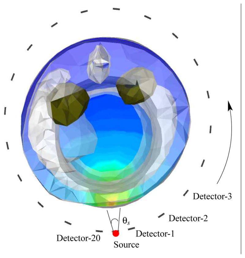
Figure 4.

Simulation setup for validating the last collided detector flux integration approach. The kidneys shown in green shade were simulated to have the uptake of 1 μM ICG. θs: the source aperture was 20 degrees. Detectors were placed 18 degrees apart.
