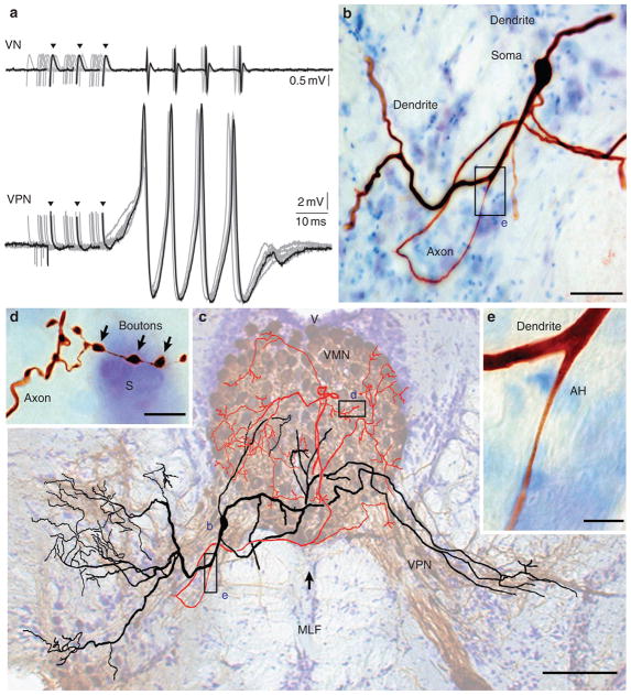
Figure 2. Vocal pacemaker neuron morphology.
(a) Intracellular vocal pacemaker neuron (VPN, bottom) and corresponding vocal nerve (VN, top) records superimposed with one highlighted (black). All records aligned to first electrically evoked VN waveform. Arrowheads indicate stimulus artefact. (b) Photomicrograph of soma, primary dendrites and axon of neurobiotin-filled VPN neuron. (c) Camera lucida drawing in transverse plane of reconstructed VPN neuron shown in (b). For general context, drawing is superimposed on background image of paired vocal motor nuclei (VMN) (arrow, midline) and VPNs bilaterally filled via transneuronal biocytin transport (brown, cresyl violet counterstain). Axonal (red) and dendritic arbors (black) overlap. (d) Photomicrograph of synaptic boutons in in VMN (S, soma) and (e) of axon hillock (AH) emerging from primary dendrite. Sites shown in (b, d, e) indicated in c. Scale bars represent 50 μm in (b), 100 μm in (c), 10 μm in (d) and 5 μm in (e). MLF, medial longitudinal fasciculi; V, fourth ventricle.

