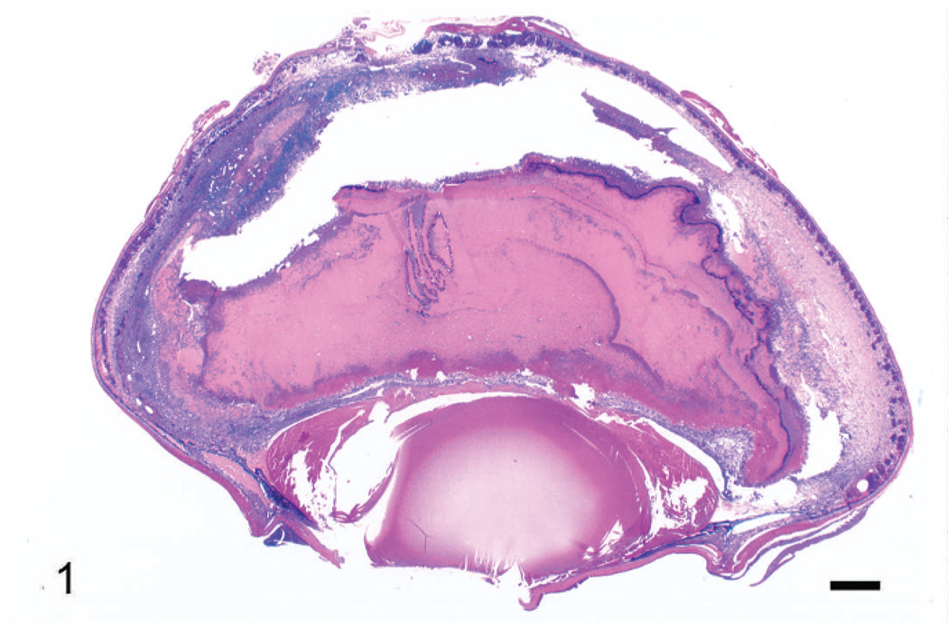
Fig. 1.
Right eye. Note retinal detachment, filling of subretinal space and vitreal chambers with amorphous eosinophilic material containing a cellular infiltrate and necrotic debris, and anterior (partially caused by collapse of the anterior chamber) and posterior synechiae. Wright’s stain. Bar = 500 µm.

