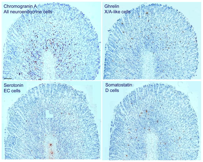
Fig. 1.
Immunohistochemical picture of neuroendocrine cells in the rat gastric oxyntic mucosa of ad libitum fed male rats. While the majority of neuroendocrine cells reside in the lower part of gastric glands, ghrelin positive X/A-like cells are evenly distributed throughout the entire length of the glands. Somatostatin-positive D cells are mostly localized in the lower half of the oxyntic glands. Serotonin-positive EC cells are rare. Scale bar represents 100 μm.

