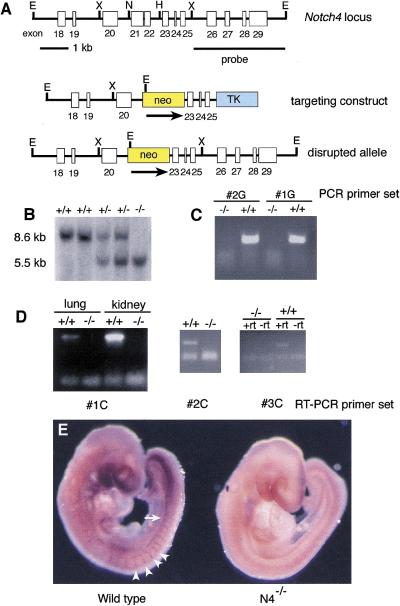
Figure 1.
Targeted disruption of the Notch4 gene. (A) Targeting scheme. (Top) The genomic organization of a portion of the Notch4 gene. Exons are indicated by white boxes. (Middle) The structure of the targeting vector. The deleted exons 21 and 22 encode amino acids 1249–1434 in the extracellular domain of the Notch4 protein. (Bottom) The predicted structure of the Notch4 locus following homologous recombination of the targeting vector. (E) EcoRI; (H) HindIII; (N) NcoI; (X) XbaI. (B) DNA isolated from embryos of the intercross of Notch4+/− heterozygous mice was digested with EcoRI, blotted, and hybridized with the indicated probe. Sizes of hybridizing fragments are indicated. Genotypes of progeny are indicated at the top of the lane. (C) Genomic PCR analysis with primers located in region deleted in the Notch4d1 mutant allele. Genotypes are indicated at top. (D) RT–PCR analysis. RT–PCR primer sets are indicated at the bottom of each panel. Primer set #1C flanks the deleted region; primers sets #2C and #3C are located at the 3′ end of the Notch4 cDNA. Genotypes are indicated at the top of the lane. (+rt) Plus reverse transcriptase; (−rt) without reverse transcriptase. (E) Whole mount in situ hybridization of a wild-type and a Notch4−/− embryo with an antisense Notch4 riboprobe encoding the intracellular domain of the Notch4 protein. Notch4 expression in the wild-type embryo is observed in intersomitic blood vessels (arrowheads) and the dorsal aorta (arrow). No Notch4 expression is observed in the mutant embryo.

