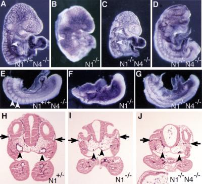
Figure 5.
Defects in vascular remodeling in Notch1−/− and Notch1−/− Notch4−/− mutant embryos. (A–G) PECAM-1-stained whole mount embryos. (A–D) Defective morphogenesis of the main trunk of the anterior cardinal vein (arrowhead) in Notch1−/− (B) and Notch1−/− Notch4−/− mutant embryos (C,D). The double mutant embryo in D is more severely affected than the embryo in C. (E–G) In the Notch1+/− Notch4−/− control embryo (E), intersomitic vessels (arrowheads) differentiate, whereas these vessels are not observed in the Notch1−/− (F) and Notch1−/− Notch4−/− (G) mutant embryos. (H–J) Histological sections of PECAM-1-stained embryos at the level of the otic vesicle. In the N1+/− control embryo (H), both the dorsal aortae (arrowheads) and the anterior cardinal veins (arrows) have open lumens and normal morphology. In the Notch1−/− Notch4−/− mutant embryo (J), endothelial cells have differentiated but both the dorsal aortae and the anterior cardinal veins have an abnormal, collapsed morphology. In the less-severely affected Notch1−/− mutant embryo (I), the anterior cardinal veins still have an open lumen but the dorsal aortae are collapsed.

