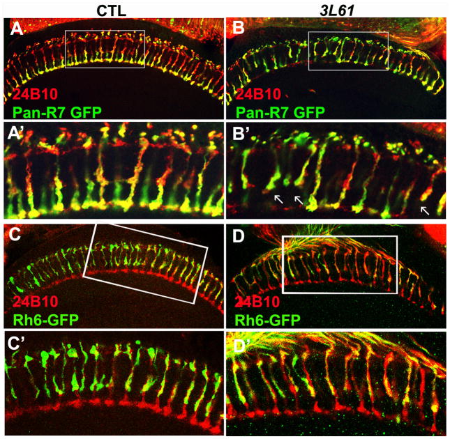
Figure 2. The R7 cells exhibit targeting defects in the 3L6 mutant animals.
(A, B, A′, B′) A portion (19.7±3%, n=268) of the R7 cells in the 3L61 mutant animals fail to target to the M6 layers but instead terminate at the M3 layers. The R7 cells were labeled with pan-R7 Gal4 that drives UAS-SytGFP (green). The arrows indicate the mistargeted R7s. (C, D, C′, D′) The R8 cells have intact target patterns. The R8 cells were labeled with Rh6-GFP (green), the photoreceptor cells were labeled with 24B10 staining (red). Panels (A′ –D′) are the enlarged views of the boxed regions in panel (A –D). The genotypes of flies used in this figure: Control (A, A′) (y w eyFLP GMRlacZ; PanR7 Gal4/UAS-SytGFP; FRT80B iso/FRT80B cl); 3L61 (B, B′) (y w eyFLP GMRlacZ; PanR7 Gal4/UAS-SytGFP; FRT80B 3L61/FRT80B cl); Control (C, C′) (y w eyFLP GMRlacZ; Rh6GFP/CyO; FRT80B iso/FRT80B cl). 3L61 (D, D′) (y w eyFLP GMRlacZ; Rh6GFP/CyO; FRT80B 3L61/FRT80B cl). (See also Figure S2)

