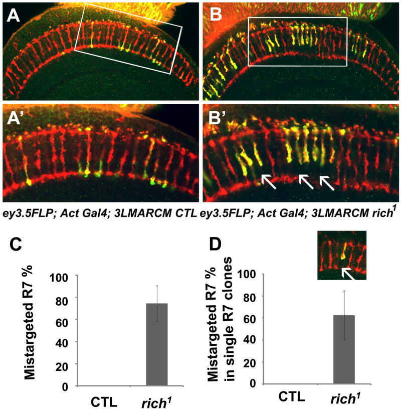
Figure 5. rich is required for synaptic specificity of R7.
Single optic section taken through a medulla of either (A, A′) control (ey3.5FLP; Act>Gal4 UAS-SytGFP/UAS-SytGFP; FRT80B M (3), tub-Gal80/FRT80B iso) or (B, B′) rich mutant (ey3.5FLP; Act>Gal4 UAS-SytGFP/UAS-SytGFP; FRT80B M (3), tub-Gal80/FRT80B rich1) animals. (A′, B′) are the enlarged images of the boxed region in (A, B). Many of the mutant R7 cells fail to target to the M6 layers (arrows). (C) Quantification of the R7 targeting defects in the ey3.5 animals (0 in control n=159;74±16% in rich n=175) illustrated in A and B. (D) 62.5 ±22% (n=37) R7 cells fail to target to the correct layer even when they are surrounded by the wild-type R7 and R8 neighbors. The small inset illustrates the single R7 mutant clone (see also Figure S4).

