Figure 6.
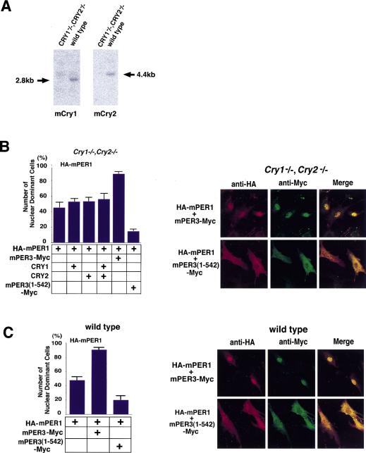
Nuclear entry of mPER1/mPER3 in mCry1/mCry2 double knockout cells. The effect of mPER3 on nuclear entry of mPER1 was investigated in mCry1mCry2 double knockout cells. (A) Northern blot analysis of mCry1 (left) and mCry2 (right) MEFs from mCry1/mCry2 double knockout mouse and wild type. In the mCry1/mCry2 double knockout cells, no signals are seen at the appropriate molecular weight. (B) Analysis of nuclear translocation of mPER1 in mCry1/mCry2 double knockout cells after coexpression of mPER3, CRY1, CRY2, and/or mPER3 (1–542). The percentage of nuclear-positive cells is shown in the bar graph. About one-half of the cells (45%) show nuclear localization by single HA-mPER1 expression, perhaps due to partnering with endogenous proteins. Although transfection of CRY1/2 did not affect the nuclear entry of mPER1, cotransfection of mPER3 markedly increased nuclear entry of mPER1. Truncated mPER3 lacking the carboxy-terminal half including the NLS prevented the nuclear accumulation of mPER1. Confocal laser microscopic images are presented at right. (C) Control nuclear translocation experiments in MEFs originating from wild-type mice expressing endogenous mCry1 and mCry2. Note the very similar nuclear localization in all combinations in C, suggesting that mCRY proteins are not essential for nuclear translocation of mPER1. All results are the mean (+s.e.m.) of three independent experiments in B and C.

