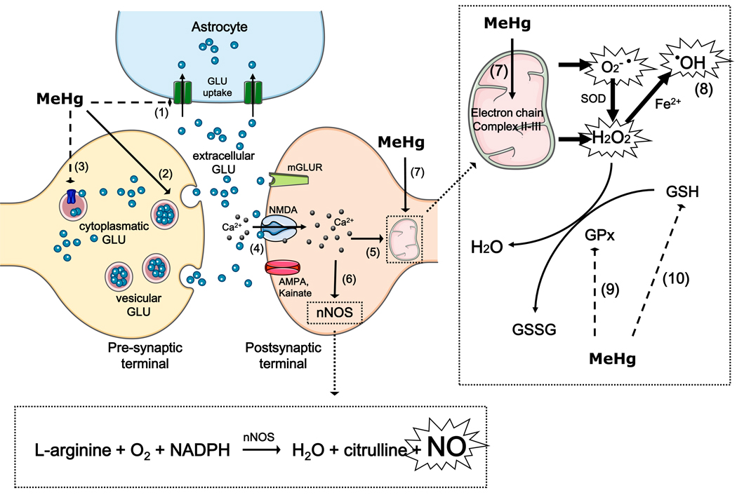
Figure 2. Reactive species as mediators of MeHg induced neurotoxicity.
MeHg leads to increased extracellular glutamate (GLU) levels through the inhibition of astrocytic glutamate uptake (event 1), the stimulation of glutamate release from presynaptic terminals (event 2) and the inhibition of vesicular glutamate uptake (event 3). Increased extracellular glutamate levels overactivate N-methyl D-aspartate (NMDA)-type glutamate receptors, increasing calcium influx into neurons (event 4). Increased levels of intracellular calcium, which can lead to mitochondrial collapse (event 5), activate neuronal nitric oxide synthase (nNOS) (event 6), thus increasing nitric oxide (NO) formation. MeHg affects the mitochondrial electron transfer chain (mainly at the level of complex II-III) (event 7), leading to the increased formation of superoxide anion (O2•−) and hydrogen peroxide (H2O2). H2O2 can produce hydroxyl radical anion (•OH) via Fenton’s Reaction (event 8). MeHg-induced increases in H2O2 levels might be a consequence of decreased glutathione peroxidase (GPx) activity (event 9) and glutathione (GSH) depletion (event 10).

