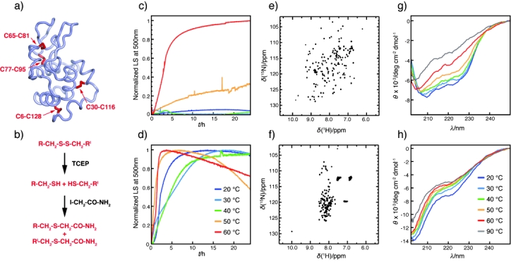
Figure 1.

a) Structure (pdb code 1Lz1) of wild-type lysozyme (Lys) with the disulfide bonds shown in red. These were reduced as shown in (b) to obtain LysRA. c, d) Amyloid formation by Lys (c) and LysRA (d) monitored by light scattering (LS) at 500 nm and different temperatures. e, f) 1H–15N NMR HSQC spectra show that e) Lys is folded whereas f) LysRA is disordered. g, h) Effect of temperature on the far-UV CD spectrum of Lys (g) and LysRA (h). TCEP=tris(2-carboxyethyl)phosphine hydrochloride.
