
Figure 7.
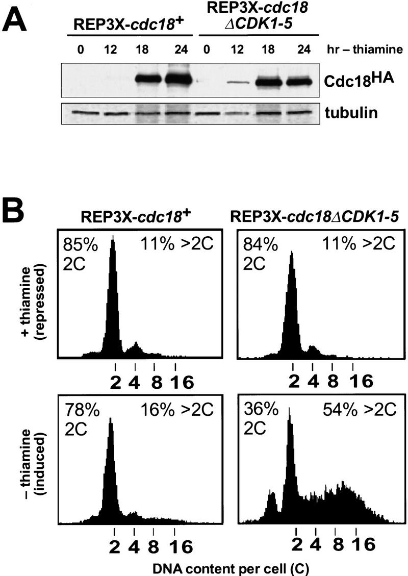


Increased replication activity associated with p65cdc18ΔCDK1-5. (A) Expression of HA-tagged cdc18+ (lanes 1–4) or cdc18ΔCDK1-5 (lanes 5–8) from the full-strength nmt1+ promoter (REP3X) was induced by growth in thiamine-free medium for the indicated times. Cell extracts were separated by SDS-PAGE and immunoblotted with monoclonal antibodies to HA and tubulin. Quantitation of p65cdc18 abundance as in Fig. 5B demonstrated that the wild-type protein accumulated to the same or slightly higher level as the ΔCDK mutant polypeptide by 18 hr. (B) The same strains were grown in the presence (upper panels) or absence of thiamine (lower panels) for 30 hr. DNA content per cell was measured by flow cytometry and is presented on a logarithmic scale. The percent of cells with normal (2C) and over-replicated (>2C) DNA content is indicated. (C) Cells in B were examined by DAPI staining of nuclei and fluorescence microscopy. The fields shown illustrate qualitatively the range of phenotypes observed in several independent experiments. When grown in the presence of thiamine, cells containing REP3X–cdc18ΔCDK1-5 were indistinguishable from those containing REP3X–cdc18+ (data not shown). (D) Wild-type cells transformed with the indicated plasmids were streaked onto EMM plates either with (+) or without (−) thiamine and incubated for 4 days at 30°C.