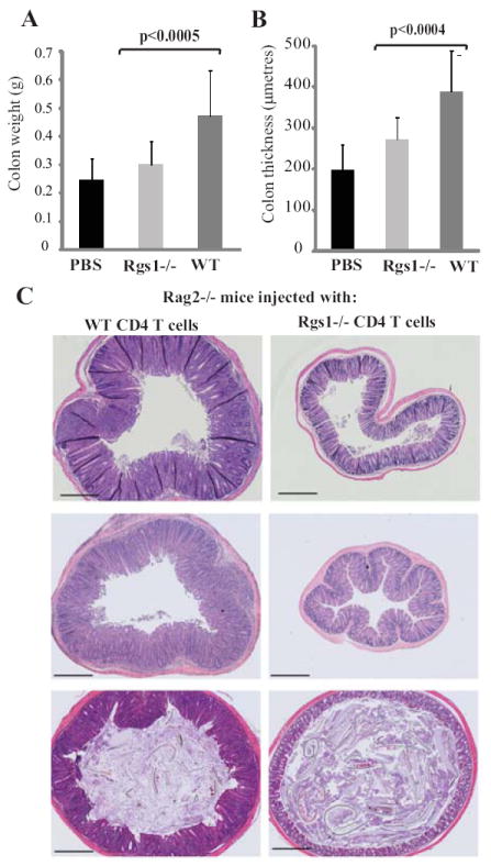
Fig 4.

Rag2-/- mice were injected in three separate experiments with either PBS (n=6) or CD4+ CD45RBhi T cells extracted from WT (n=13) or Rgs1-/- mice (n=11). Colons were removed 7-8 weeks later, weighed and sectioned. Mean colon weight (A) and medial colon thickness (B) in recipients of WT (dark grey bars) or Rgs1-/- T cells (light grey bars) or PBS (black bars): statistics by Student’s T test. (C) Representative mid colon histologies at 7 weeks for recipients of WT (left) or Rgs1-/- cells (right). One colon for each group is shown from each of three independent experiments. Scale bar =500μm.
