
Figure 2.
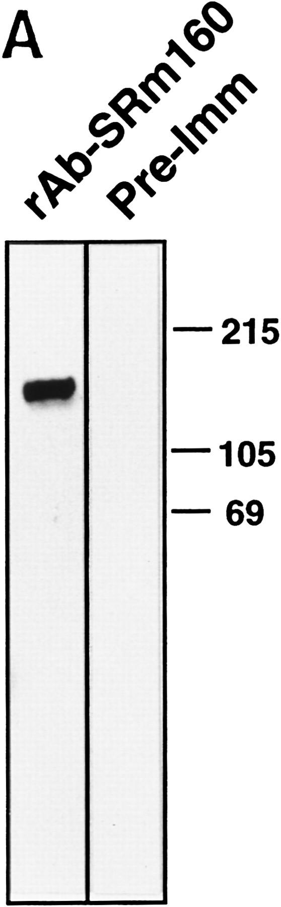
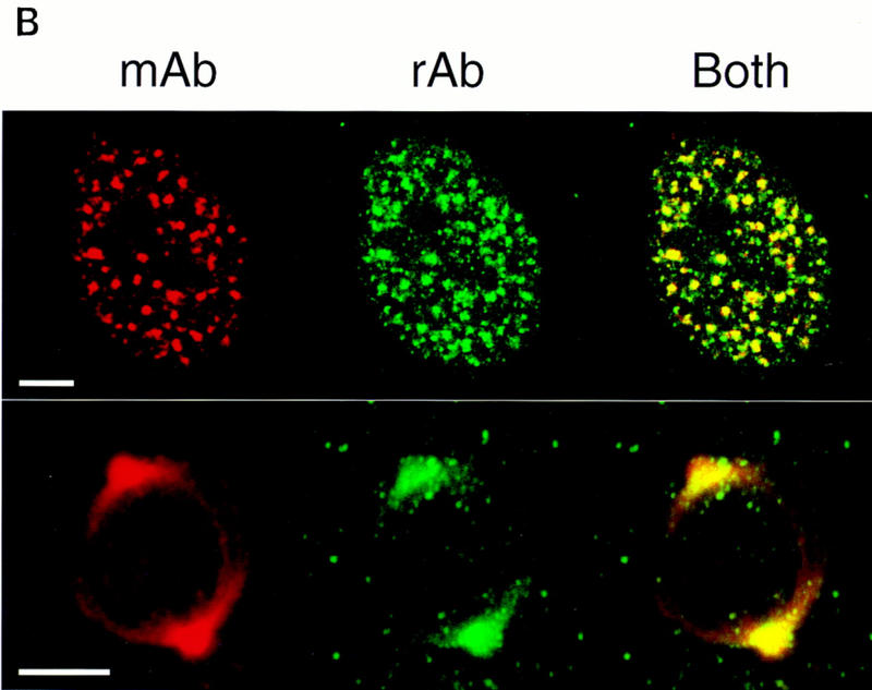
Association of SRm160 with interphase nucleoplasmic speckles, foci, and the mitotic spindle apparatus. (A) Total HeLa nuclear extract was separated on an SDS–polyacrylamide gel and immunoblotted with an affinity-purified polyclonal antiserum raised to a GST–SRm160 fusion protein containing SRm160 ORF amino acids 7–160 (rAb–SRm160, lane 1), and the corresponding preimmune serum (lane 2). (B) Interphase (top row) and metaphase (bottom row) human CaSki cells were double-immunolabeled with mAb B1C8 (red) and rAb–SRm160 (green) and visualized by confocal microscopy. The images were superimposed to reveal sites of overlap (yellow). The interphase image corresponds to a single confocal section proximal to the midline of the nucleus. The metaphase image corresponds to a stack of merged sections through the whole cell. Bar, 5 μm.