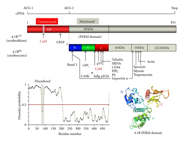
Figure 4.

Primary structure of 4.1R isoforms and map of known binding partners for 4.1R. Translation of the prototypical red blood cell 80 kDa 4.1R isoform (4.1R80) is initiated at AUG-2, which is located in exon 4. Translation of the 135 kDa 4.1R isoform (4.1R135), an isoform expressed in early erythroblasts and other nucleated cells, is initiated at AUG-1, which is located in exon 2′ (ID: P11171). The 30 kDa membrane-binding domain is the so-called “FERM” domain. Disorder prediction for each domain has been established through the use of the PrDOS software package. An updated list of the binding partners identified for each domain of 4.1R is displayed. CPAP refers to a “centrosomal protein 4.1R-associated protein” reported by Hung et al. [24]. A 3D representation of the 30 kDa FERM domain of 4.1R, visualized with the MolFeat Ver. 4.6 software, is displayed (PDB accession no. 1GG3). The 30 kDa domain consists of three lobes (N-, α-, and C-lobe) and adopts a three-leaf clover shape [25].
