
Figure 4.
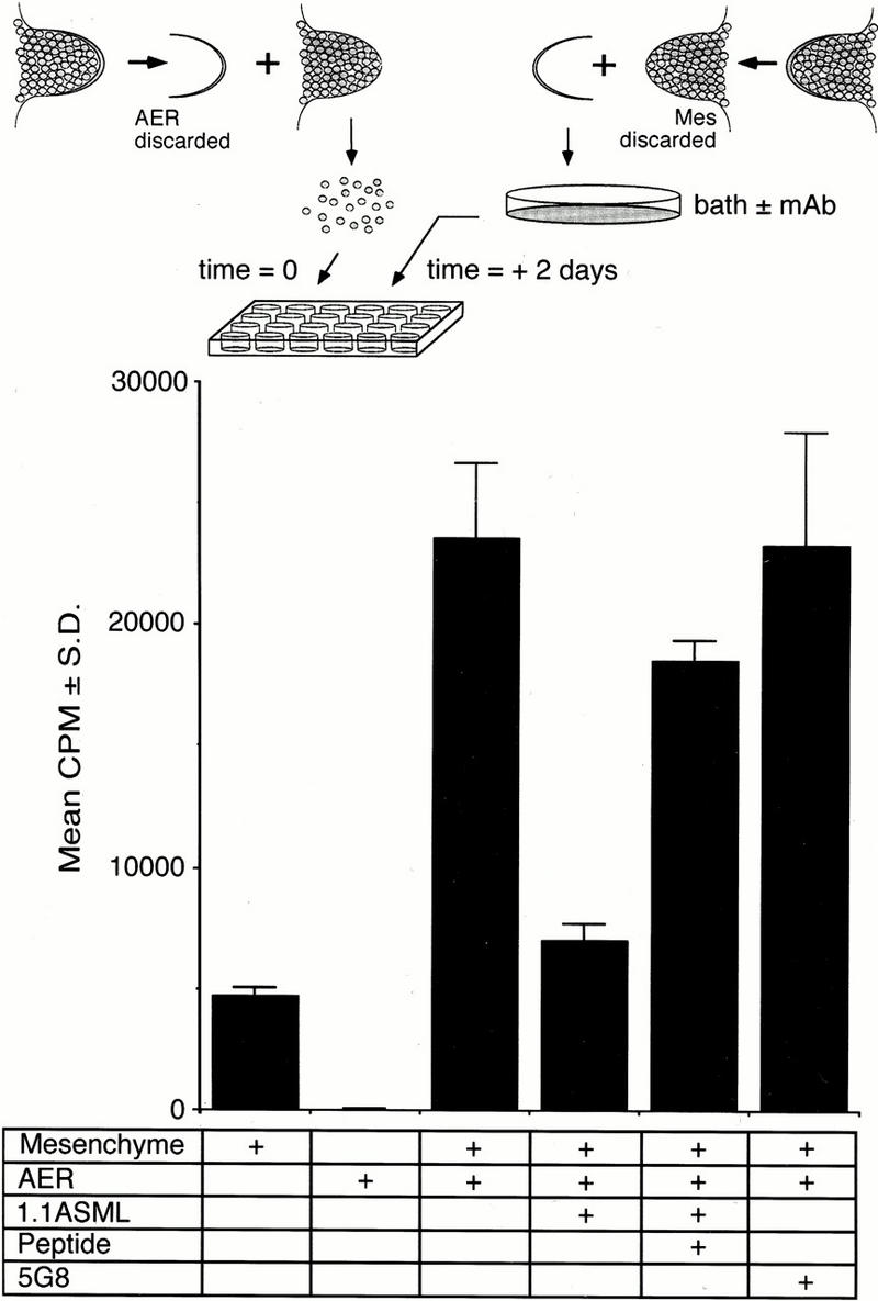
AER-mediated proliferation of primary mesenchymal cells in culture. Limb bud mesenchymal cells were put into culture as described in Materials and Methods. AER fragments were independently collected, treated either in PBS or in PBS containing 5G8, 1.1ASML, or 1.1ASML preblocked by its recognition peptide, washed, and placed on top of the mesenchymal cells. Within 2–3 hr the AER made adhesive contact to the mesenchymal cell layer. After 16 hr, thymidine incorporation was determined as a measure of mesenchymal cell proliferation. Plotted cpm represent the mean of triplicate samples ± s.d.