
Figure 6.
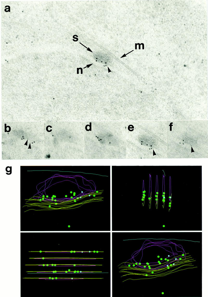
Electron microscopic localization of Cut12 protein within the SPB. (a) Immunogold staining of a section through an SPB by use of antibodies to the Pk epitope tag and a polyclonal anti-Cut12 antibody. Secondary antibodies were conjugated to gold particles of 5 nm (Pk) and 10 nm (Cut12). (S) Spindle pole body; (M) cytoplasmic microtubule laterally associated with the SPB; (N) nuclear envelope. The Cut12 epitopes were localized specifically to the inner face of the main body of the cytoplasmic SPB, adjacent to the nuclear envelope. The Pk epitope tag appeared localized more to one side of the inner face of the SPB. (b–f) Five serial sections through the single SPB shown in a were stained with antibodies to the Pk epitope tag and polyclonal antibodies to the Cut12 protein. Secondary antibodies were conjugated to gold particles of 5 nm (Pk) and 10 nm (Cut12). (g) Immunogold localization of Cut12 epitopes in serial sections through a single SPB in the Pk epitope tagged cut12+ strain. The five serial sections shown in b–f were superimposed. (Green dots) 10 nm-gold particles (anti-Cut12); (blue dots) 5 nm gold particles (anti-Pk); (yellow lines) nuclear envelope; (red lines) SPB; (blue line) cytoplasmic microtubule.