
Figure 2.
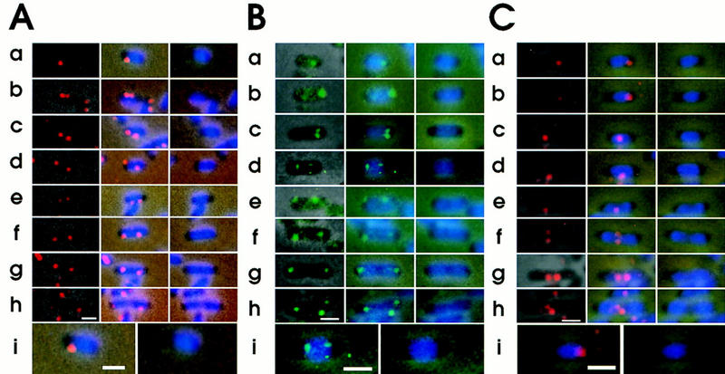
Intracellular localization of replication origin and terminus detected by FISH. Cells of strain CSH50, grown exponentially at 37°C in M9 minimal glucose medium, were fixed. The fixed cells were hybridized with the Cy3-labeled oriC–gid DNA probe (A), the fluorescein-labeled asn–rbs DNA probe (B), and the Cy3-labeled ter DNA probe (C). (Left columns) Combined images of the phase contrast micrograph and fluorescence micrograph for FISH; (middle columns) combined images of the phase contrast micrograph, fluorescence micrograph for FISH and fluorescence micrograph for DAPI; (right columns) combined images of the phase contrast micrograph, and fluorescence micrograph for DAPI. The cells from A row a, B row d, and C row a are shown at higher magnification. Bar, 1 μm.