
Figure 5.
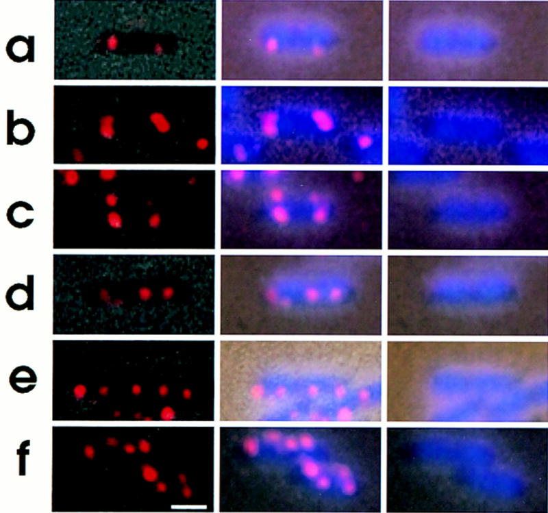
Intracellular localization of replication origin. Cells were grown, fixed and hybridized with the oriC–gid DNA probe as described in the legend to Fig. 2. (Left column) Combined images of the phase contrast micrograph and fluorescence micrograph of the same field; (middle column) combined images of fluorescence micrograph and phase contrast-DAPI micrograph; (right column) images of phase contrast-DAPI. Bar, 1 μm.