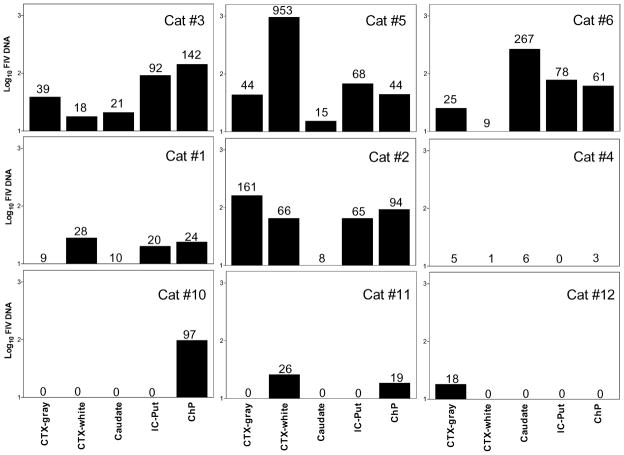
Figure 6.
FIV proviral DNA in various brain regions of individual i.c.v. cats (cats 1 to 6) or i.p. cats (cats 10 to 12). Total genomic DNA was extracted from 30 mg tissue pieces of four brain regions, and the choroid plexus. FIV proviral DNA was quantified by real-time PCR and the proviral loads were expressed as FIV copies per microgram total genomic DNA. FIV DNA was detected in all the i.c.v. cats although the regional levels of proviral DNA were quite variable between individual cats. The number of FIV copies/μg genomic DNA detected is indicated above each bar. The limit of detection for the PCR was approximately 10 copy equivalents/μg genomic DNA and values at or below this level are show numerically with no associated bar. The overall proviral loads in the four brain regions are substantially higher and more widespread in i.c.v. cats than i.p. cats. Regions sampled: frontal cortical gray matter, CTX-gray; frontal cortex white matter immediately under the gray matter, CTX-white; caudate nucleus, internal capsule–putamen, IC-Put; and choroid plexus, ChP.

