Fig. 3.
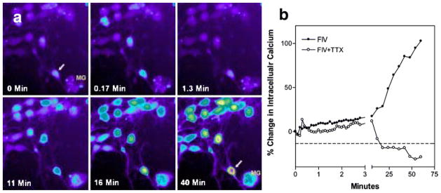
Example of intracellular calcium changes in cultured feline neurons isolated from fetal cortex–hippocampus. (a) A typical cluster of neurons is shown with low resting calcium at 0 min. The intensity of the Fluo-3 fluorescence is proportional to the cytosolic calcium concentration and is color-coded from low to high as: purple>blue>green>yellow>red>white. Addition of FIV isolated from the CSF of an infected cat resulted in a small transient increase in a few neurons (0.17 min), followed by a gradual increase beginning as early as 11 min. Moderately high calcium levels developed in many neurons by 40 min. Individual neuronal responses can be quite variable, ranging from minimal responsiveness to robust increases as seen in an isolated neuron (arrow) adjacent to a microglial cell (MG). (b) Effect of synaptic activity on the calcium rise can be seen in a comparison between neurons exposed to FIV in the presence or absence of 1 μM tetrodotoxin (TTX). A small acute increase is seen in both preparations but the TTX-treated neurons failed to show the delayed calcium deregulation, indicating that synaptic activity is necessary for the delayed calcium rise.

