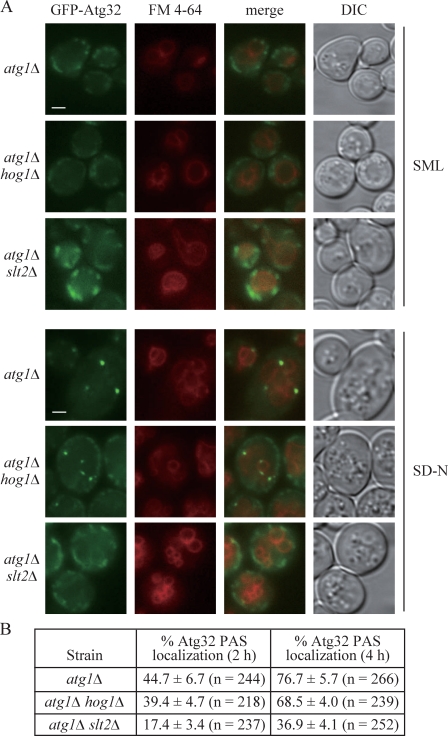
Figure 6.
Recruitment of Atg32 to the PAS is defective in the slt2Δ, but not the hog1Δ mutant. (A) Plasmid-driven GFP-Atg32 was transformed into atg1Δ (WHY001), atg1Δ slt2Δ (KDM1203), and atg1Δ hog1Δ (KDM1211) strains. Cells were grown to mid-log phase in SML, shifted to SD-N for 2 or 4 h, and stained with FM 4-64 to mark the vacuole. Representative pictures from single Z-section images are shown. DIC, differential interference contrast. Bars, 2.5 µm. (B) Quantification of Atg32 PAS localization. 12 Z-section images were projected and the percentage of cells that contained at least one GFP-Atg32 dot on the surface of the vacuole was determined. The SD was calculated from three independent experiments.

