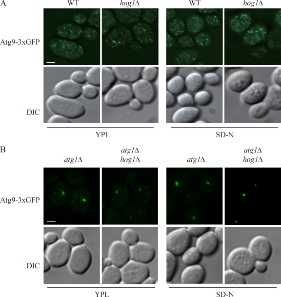
Figure 7.
Atg9 movement is unaffected in the hog1Δ mutant. (A and B) The localization of Atg9-3xGFP was tested in wild-type (JGY134) and hog1Δ (KDM1207) strains (A) and atg1Δ (JGY135) and atg1Δ hog1Δ (KDM1212) strains (B). Cells were cultured in growing (YPL medium) conditions and shifted to nitrogen starvation medium (SD-N) for 2 h. Cells were fixed and observed by fluorescence microscopy. Representative pictures from single Z-section images are shown. DIC, differential interference contrast. Bars, 2.5 µm.

