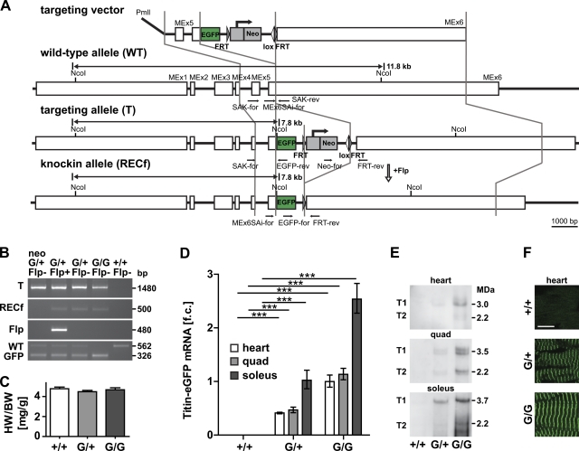
Figure 1.
Generation of the titin-eGFP knockin mouse. (A) Targeting strategy to insert eGFP into titin’s M-band exon 6 (MEx6). The targeting vector was linearized with PmlI and contains the FRT flanked Neo selectable marker. Locations of the genotyping primers are indicated in the wild-type, targeted, and knockin allele (black arrows). Germline expression of the Flp recombinase was used to excise the Neo cassette (open arrow). (B) PCR-based genotyping of wild-type (+/+), heterozygous (G/+), and homozygous (G/G) titin-eGFP knockin mice. The targeted allele (T) and the excision of the Neo cassette (RECf allele) after introduction of the Flp transgene (Flp) were confirmed by PCR. Homozygous knockin animals are viable as documented by the absence of the wild-type allele in the GFP-positive animal (G/G Flp−). Bp, base pair. (C) Heart to body weight ratio of female heterozygous (G/+) and homozygous (G/G) titin-eGFP knockin mice was unchanged compared with wild-type (+/+; n = 6 per group). (D) Expression analysis of titin-eGFP in heart and skeletal muscle (quadriceps and soleus) using qRT-PCR confirmed the expected intermediate titin-eGFP mRNA levels in heterozygous (G/+) versus homozygous animals (G/G). Soleus muscle mRNA levels were increased as compared with heart and quadriceps. Data were normalized to 18S RNA and titin levels in homozygous hearts were set as 1 (n = 5 per group). f.c., fold change. ***, P < 0.001. (E) Western blot using an anti-GFP antibody detects the high molecular weight titin-eGFP fusion protein (T1, full length; T2, degradation product). (F) The eGFP-tagged titin protein was integrated into the sarcomere as indicated by confocal imaging of native cardiac tissue. In both hetero- and homozygotes, the fluorescent signal was sufficiently strong for detection without amplification. Bar, 10 µm. Error bars indicate SEM.

