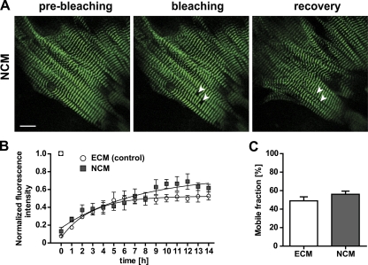
Figure 8.
Titin-eGFP dynamics in neonatal cardiomyocytes. (A) FRAP was completed within 14 h in neonatal cardiomyocytes (NCM) prepared on postnatal day two (P2). White arrows indicate the bleached regions. Bar, 10 µm. (B) Comparison of embryonic vs. neonatal cardiomyocytes (ECM E13.5 vs. NCM P2) did not reveal a significant difference (ECM, n = 4; NCM, n = 3). (C) There was no difference in mobile fractions between neonatal and embryonic cardiomyocytes (ECM, n = 4; NCM, n = 3). Error bars indicate SEM.

