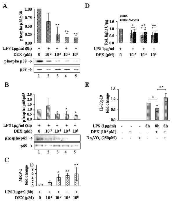
Figure 4.
DEX down-modulates p38 MAPK activity. (A) DEX-dependent inhibition of p38 MAPK was measured in LPS-stimulated Mφ by western immunoblotting of the phosphorylated protein (phospho Thr180/Tyr182), normalized on the total amount of p38. Data are reported in bar graph as fold changes relative to the DEX-untreated sample (lane 1); n = 3. (B) Inhibition of NF-κB-p65 transactivity by DEX was measured in LPS-stimulated Mφ by western immunoblotting of p65 phosphorylation on Ser276, normalized on the total amount of p65. Data are reported in bar graph as fold changes relative to the DEX-untreated sample (lane 1); n = 3. (C) DEX-induced increase of MKP-1 mRNA determined by RT-PCR on the same samples as in (A). Data are reported as fold changes relative to the DEX-untreated sample (lane 1); n = 6. (D) Bar graph shows data from luciferase assays performed on MM6 reporter cells, stimulated with LPS and treated with increasing concentrations of DEX, administered alone or with sodium orthovanadate. Data are reported as fold changes relative to the DEX-untreated sample (+LPS, 0 DEX); n = 15. (E) Bar graph shows RT-PCR data for the IL-23p19 transcript from Mφ stimulated with LPS and then treated or not with DEX 0.01 μM alone or along with sodium orthovanadate. Data are reported as fold changes relative to the DEX-untreated sample (8 hLPS, - DEX, -Na3VO4); n = 6. * p < 0.05, ** p < 0.01.

