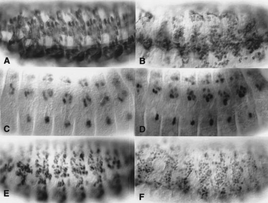
Figure 2.
MHC, NAU, and MEF2 positive cells are present in sns mutant embryos but do not fuse to form muscle fibers. All embryos are oriented ventrolaterally with anterior to the left. Wild-type embryos are shown in A, C, and E. (B,D,F) Embryos that are genetically snsA3.24/Df(2R)BB1. Embryos homozygous for snsA3.24 exhibit the same mutant phenotype (data not shown) and Df(2R)BB1 completely removes the sns genomic region (Results, Materials and Methods). Therefore, by genetic criteria, snsA3.24 appears to represent the null phenotype for sns. (A,B) Stage 15 embryos immunostained with an MHC antibody to visualize the musculature. By comparison with wild-type in A, embryos mutant for sns exhibit unfused MHC expressing myoblasts in the place of mature muscle fibers. (C,D) Stage 13 embryos immunostained with antisera against NAU to visualize a subset of founder cells. In wild-type embryos (C), NAU-expressing cells are distributed in an array of muscle founders and precursors. (D) The pattern of NAU-expressing cells is not affected in sns mutant embryos, suggesting that founder cell specification does not require sns. (E,F) Stage 15 embryos immunostained with antisera against MEF2 to visualize the entire myoblast population. MEF2 expression can be seen in developing muscle fibers in wild-type embryos (E) and in the corresponding population of unfused myoblasts in sns mutant embryos (F).

