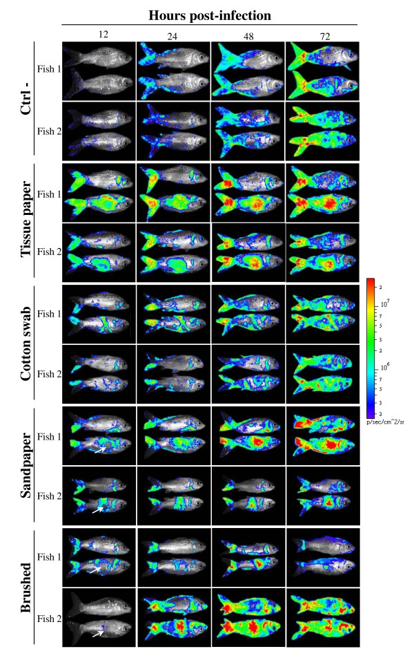
Figure 3.
Effect of skin physical treatments on CyHV-3 entry in carp analyzed by bioluminescence imaging. Each physical treatment depicted in Figure 2 was applied to a group of 7 fish. Immediately after skin treatment, fish were inoculated by immersion in water containing the FL BAC 136 LUC TK revertant strain (103 PFU/mL of water for 2 h) to mimic natural infection. The fish were analyzed by bioluminescence imaging at the indicated time post-inoculation. Each fish was analyzed lying on its right and its left side. Two representative fish are shown per group. White arrows indicate the centre of epidermis lesions which was associated with no bioluminescent signal at 12 h post-inoculation but an intense signal later during infection. The images collected over the course of the experiment are presented with standardized minimum and maximum threshold values for photon flux.

