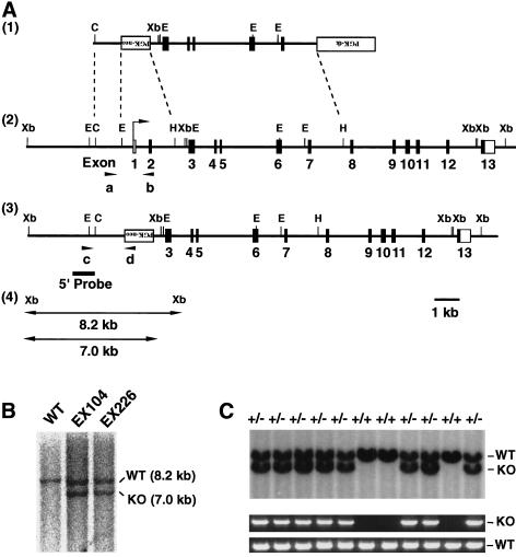
Figure 1.
Targeted disruption of mouse Chk1. (A) Structure of the targeting vector pVT–Chk1N (1), restriction map of the mouse Chk1 locus (2), and structure of the mutated locus after homologous recombination (3). The coding and noncoding exons are numbered and depicted by closed and open boxes, respectively. HindIII sites other than those shown also exist in the locus. The mutated Chk1 allele was detected by PCR with a set of primers, c and d, shown as arrowheads, and by Southern blot analysis, with a 5′ probe shown as a bold line. The expected sizes of the XbaI fragments that hybridize with the probe on Southern analysis are indicated for the wild-type and mutant Chk1 alleles (4). (PGK–neo) Neomycin transferase gene linked to the phosphoglycerate kinase (PGK) gene promoter; (PGK–tk) thymidine kinase gene of herpes simplex virus linked to the PGK gene promoter; (B) BamHI; (E) EcoRI; (C) ClaI; (Xb) XbaI; (H) HindIII. (B) Southern blot analysis of genomic DNA from targeted ES clones EX104 and EX226. Genomic DNA from individual ES clones was digested with XbaI and hybridized with the 5′ probe. The 8.2-kb fragment corresponding to the wild-type (WT) allele and the 7.0-kb fragment corresponding to the mutated (KO, knockout) allele are indicated. (C) Genotype analysis by Southern blot analysis (top) and PCR (bottom) of E11.5 embryos from heterozygote intercrosses. DNA samples were subjected to PCR with primers a and b for the wild-type (WT) allele and with primers c and d for the mutated (KO) allele, yielding amplification products of 0.9 and 1.5 kb, respectively.

