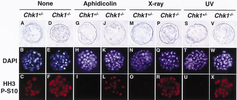
Figure 5.
Abrogated G2 checkpoints in Chk1−/− embryos. Chk1+/− and Chk1−/− blastocysts at E3.5 were either not treated (A–F) or were treated with 1 μm aphidicolin (G–L), X ray (10 Gy; M–R), or UV (0.07 J/cm2; S–X). Three hours after treatment, embryos were incubated with nocodazole (0.1 μg/ml) for an additional 3 hr and then fixed and stained with antibody specific to phosphohistone H3 at Ser10 (HH3 P-S10; C,F,I,L,O,R,U,X) and DAPI (B,E,H,K,N,Q,T,W). The genotypes of each embryo were determined by PCR. Images were obtained under either bright-field conditions or ultraviolet illumination.

