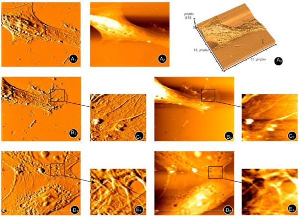
Figure 5.
Atomic force microscopy of hMSCs under normoxic conditions. A typical long spindle, cytoskeleton (A1, A2, A3), and palpus-like or cicada wing-like pseudopodium (B1,B2, enlarged areas shown in C1 and C2 respectively) were observed. The mesh-like cytoskeleton of adjacent hMSCs (D1, D2) can also been seen (enlarged areas shown in E1, E2).

