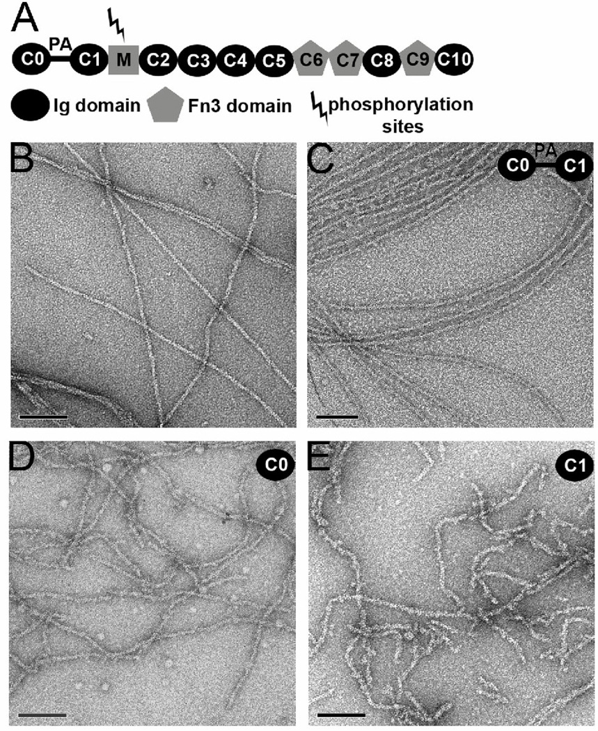
Figure 1.
The modular structure of c-MyBP-C includes multiple Ig and fibronectin domains (A). A proline/alanine rich (PA) region links C0 with C1. Electron micrographs of negatively stained F-actin (B), and F-actin decorated with C0-C1 (C), C0 alone (D) and C1 alone (E). The space bars are 1,000 Å. When C0-C1 is added to F-actin extensive cross-linking of filaments is observed (C, inset), but regions can still be found where images of single filaments can be used for three-dimensional reconstruction.

