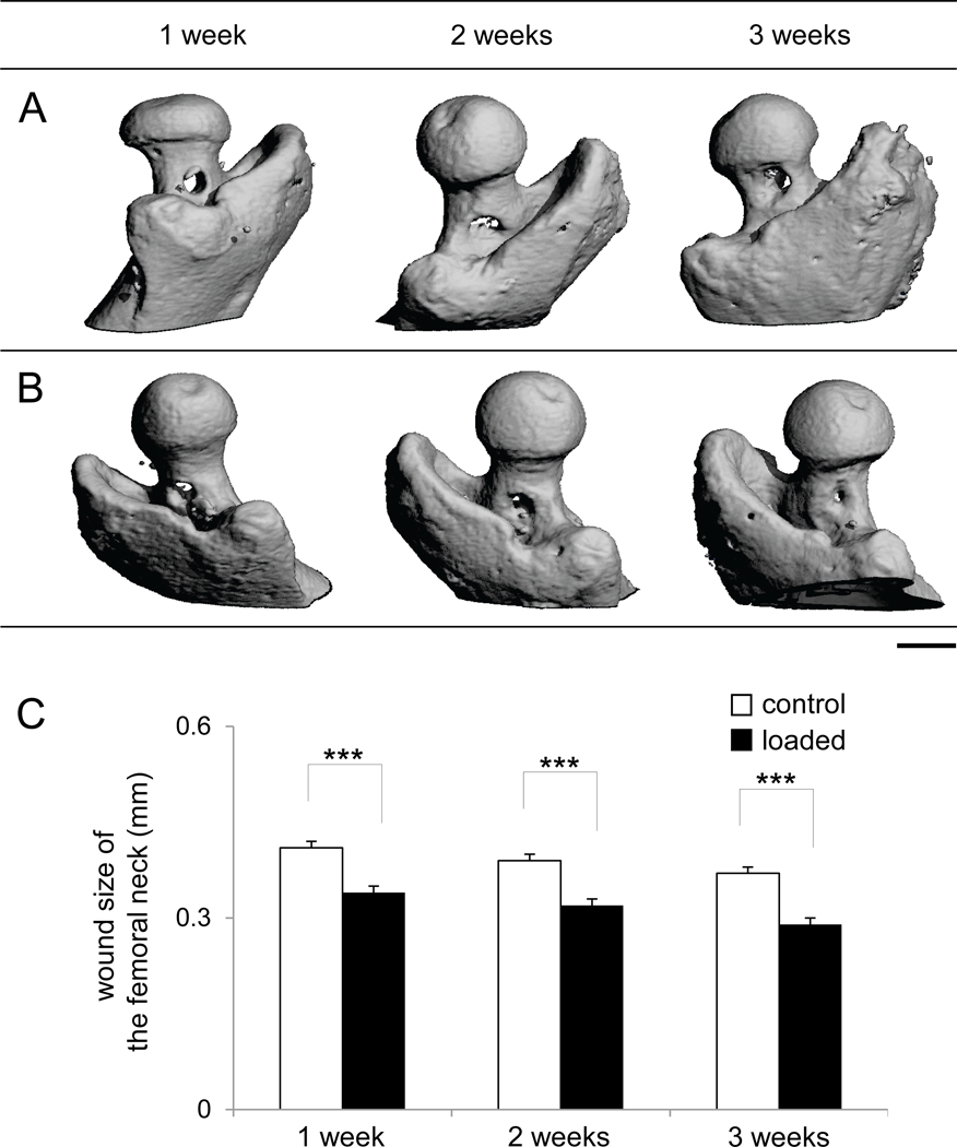
Figure 5.
Surgical wounds in the femur neck. (A) Micro CT images of the surgical holes of the control femora in the femur necks in 1, 2, and 3 weeks after surgery. (B) Micro CT images of the surgical holes of the loaded femora in the femur necks in 1, 2, and 3 weeks after surgery. Bar = 1 mm. (C) Changes in wound size in the femur neck (mm).

