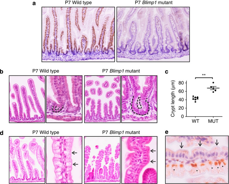
Figure 3. Accelerated maturation of Blimp1 mutant intestinal epithelium.
(a) Immunohistochemistry for Blimp1 shows efficient loss of Blimp1 from the intestinal epithelium. (b) In the proximal small intestine, there was a noticeably enhanced formation of crypts (dotted line) at P7 compared with the rudimentary crypts (dotted line) of the wild-type mice. (c) Average crypt depth in wild type (WT, n=7) and Blimp1 mutant (MUT, n=5) animals, average plus s.e. (d) In the distal small intestine of WT mice, enterocytes clearly showed the translucent cytoplasm that is typical for the presence of large cytoplasmic vacuoles that characterize this stage (arrows). These vacuoles were completely absent in Blimp1 mutant enterocytes (arrows). (e) A rare villus on which Blimp1 is only partially deleted clearly shows the difference in phenotype between Blimp proficient (brown nuclear staining, asterisks) and deficient cells (arrows). **P<0.01, Student's t-test. Original magnifications ×200 for overviews and ×800 for enlargements.

