
FIGURE 2.
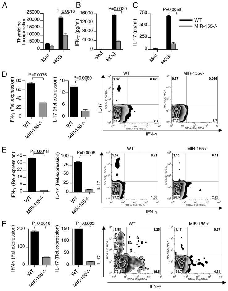
Mir-155−/− mice exhibit lower Th1 and Th17 cytokine production during EAE. Splenocytes from WT and Mir-155−/− mice were harvested 10 d after immunization and were stimulated ex vivo with MOG peptide 35–55. A, In the last 16 h, cells were pulsed with thymidine and assayed for proliferation (cpm). Error bars represent SEM between triplicates. Supernatants from parallel cultures were harvested 72 h after initiation of cultures and assayed by ELISA for IFN-γ (B) and IL-17 (C). Real-time RT-PCR analysis and intracellular staining of IFN-γ and IL-17 in (D) spleen-, (E) LN-, and (F) CNS-derived CD4+ T cells isolated from WT and Mir-155−/− mice with EAE. Data in A–F are representative of three experiments.