
Figure 2.
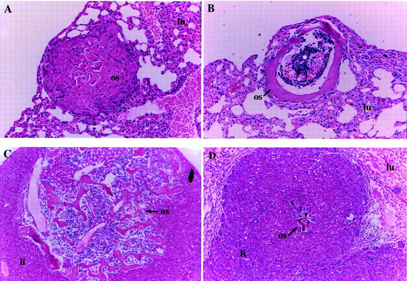
Tumors that arise in Nf2 heterozygous mice exhibit a very high rate of metastasis. (A) A fairly undifferentiated osteosarcoma metastasis (os) in the lung (lu) of a Nf2 +/− mouse. Some mineralization is present, but there is abundant cellularity. This is the typical histological appearance of an osteosarcoma metastasis (200×). (B) Highly differentiated metastasis in a Nf2 +/− mouse. This is an unusual form of metastasis frequently seen in Nf2 heterozygotes. The mineralized bone has formed a collar as in a long bone, and some lymphocytes appear to have homed to the “pseudobone marrow cavity” in the center (asterisk) (100×). (C) A highly differentiated osteosarcoma metastasis (os) in the liver (li) of a Nf2 +/− mouse. Again, there appears to be lymphocyte homing and formation of “bone marrow.” Two large blood vessels are also present within the metastasis and probably provided the route of entry for the tumor cells into the lung (100×). (D) A metastasis from a hepatocellular carcinoma (li) to the lung. This mouse also had an osteosarcoma that metastasized to the liver metastasis (os; center). The normal lung tissue is compressed in the upper corners (lu) (100×).