
Figure 5.
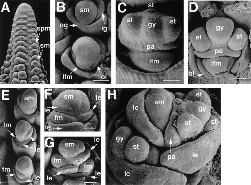
Scanning electron microscopy of normal and ids1–mum2 ears. (A) A632 ear primordium with initiating spikelet pair primordia and spikelet primordia. Bar, 300 μm. (B) A632 unbranched spikelet meristem (top) and slightly older branched spikelet meristem (bottom). The older spikelet meristem undergoes lateral branching to initiate the lower floret. Bar, 102 μm. (C) Development of lateral organs in A632 ear spikelets. The lateral organs of the upper floret develop first. Bar, 80 μm. (D) Maturing lateral organs of the upper floret of A632 ear spikelets with initiating gynoecial ridge. Bar, 104 μm. (E) Branching ids1–mum2 spikelet meristems. The uppermost spikelet meristem has not branched yet and appears elongated. Simultaneous floret branching and lemma initiation occurs on opposite sides of the middle and lower spikelet meristems. Bar, 156 μm. (F) Further development of ids1–mum2 spikelet meristems with glumes removed showing development of the floral meristems within the axils of the lemmas. The spikelet meristem persists after each branching event. Bar, 58 μm. (G) Older ids1–mum2 spikelet meristem with glumes removed. The residual spikelet meristem elongates to initiate another floret. Bar, 60 μm. (H) ids1–mum2 spikelet showing maturing lateral organs. There is no distinction between upper and lower floret in terms of lateral organ development. The youngest floret branching from the spikelet meristem is obscured by the lemma. Bar, 171 μm. (spm) Spikelet pair meristem; (sm) spikelet meristem; (lfm) lower floral meristem; (gy) gynoecium; (fm) floral meristem; (le) lemma.