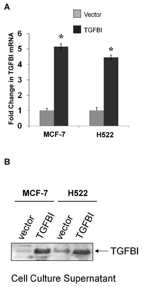
Fig.1.

Establishment of cell lines stably expressing TGFBI. MCF-7 and H522 cells were transfected with TGFBI cDNA or empty vector. The expression of exogenous mRNAs and proteins was confirmed by RT-PCR and Western blotting, respectively. (A) Relative quantification of TGFBI mRNA was determined by real-time RT-PCR using the ΔΔCt method with GAPDH as an internal control. Results are mean±SD from three independent experiments. * p<0.05 versus vector controls. (B) Expression of TGFBI in cell supernatant was determined by Western blotting. Experiments were performed three times and a representative immunoblot is shown here.
