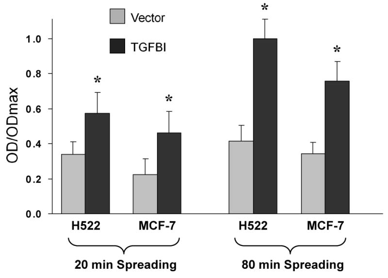
Fig.2.

Adhesion of cells expressing TGFBI to fibronectin. 1×10⁴ cells were maintained in suspension for 40 min and then allowed to adhere to fibronectin-coated plates for the indicated times. Bound cells were fixed and stained with crystal violet before optical density (OD) was measured at 595 nm. Adhesion to fibronectin was significantly enhanced after expression of TGFBI in H522 and MCF-7 cells. Data are normalized to the maximum OD detected, and expressed as mean±SD from three independent experiments. *, p<0.05 versus vector controls.
