Fig.3.


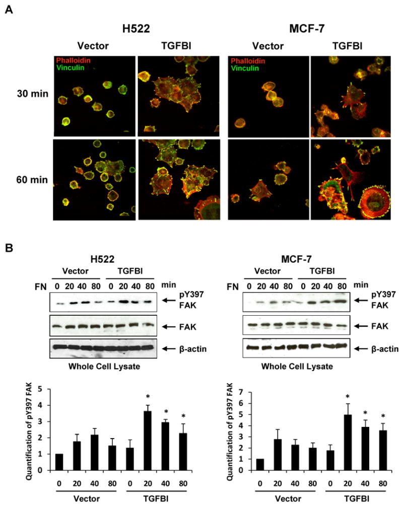
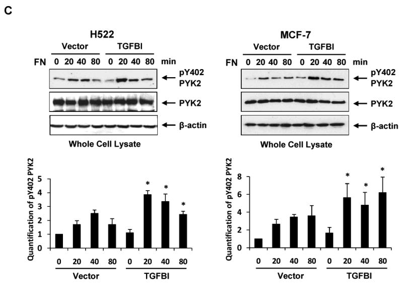
Alteration of focal adhesion and actin stress fiber formation with associated signaling activation in H522 and MCF-7 cells expressing TGFBI. (A) Representative images display focal adhesion and stress fiber formation when spread on fibronectin, which are dramatically increased in cells expressing TGFBI. Cells were plated on fibronectin-coated coverslips in DMEM containing 2% bovine serum albumin and incubated at 37°C for indicated time points. The adherent cells were then fixed and stained with rhodamine phalloidin (red) for stress fiber or antibody to vinculin (green) for focal adhesion (magnification 100×). Activation of (B) FAK and (C) Pyk2 after plating on fibronectin was greatly enhanced in the presence of TGFBI. To detect activated FAK and Pyk2, immunoblot membranes were probed with antibodies to phospho-Y397-FAK and phospho-Y402-Pyk2. Membranes were also reprobed with anti-FAK, anti-Pyk2 and anti-β-actin antibodies to demonstrate equal protein loading. The intensity of protein bands was quantified by ImageJ developed by the National Institutes of Health and plotted using MS-EXCEL. *, p<0.05 versus corresponding time point of vector controls. The figures represent one of the three independent experiments.
