
Figure 2.
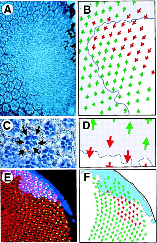
LOF and GOF JAK/STAT pathway clones show abnormal ommatidial polarity. (A) A large dorsal hop2 clone (marked by absence of white+ pigment granules) contains normally differentiated ommatidia in both correct and inverted orientation with respect to surrounding wild-type clusters. (B) Schematic representation of the clone shown in A. Dorsal-oriented ommatidia are shown as green arrows and inverted ommatidia are shown in red. The approximate position of the clonal boundary is shown by the purple line. A field of inverted ommatidia is present around the polar margin of the clone. (C) A high-magnification view of ommatidia along the polar boundary of a hop2 clone (marked by absence of white+) showing an inverted ommatidium comprised of entirely wild-type photoreceptor cells (note dark pigment granules associated with the rhabdomeres of wild-type photoreceptors, indicated by arrows). (D) Schematic representation of clone shown in C. Dorsal ommatidia are shown by green and ventral by red arrows. Boundary of the clone is indicated. (E) A clone of cells ectopically expressing Upd (blue) in the dorsal part of an eye disc stained to show Elav (red) and Prospero (green, yellow in overlay). Ommatidia with inverted orientation are seen on the ventral edge of the clone. (F) A schematic representation of clone shown in E indicating the extent of the Upd expressing clone (pale blue) and the ommatidial orientation indicated by dorsal green and ventral red arrows.