
Figure 3.
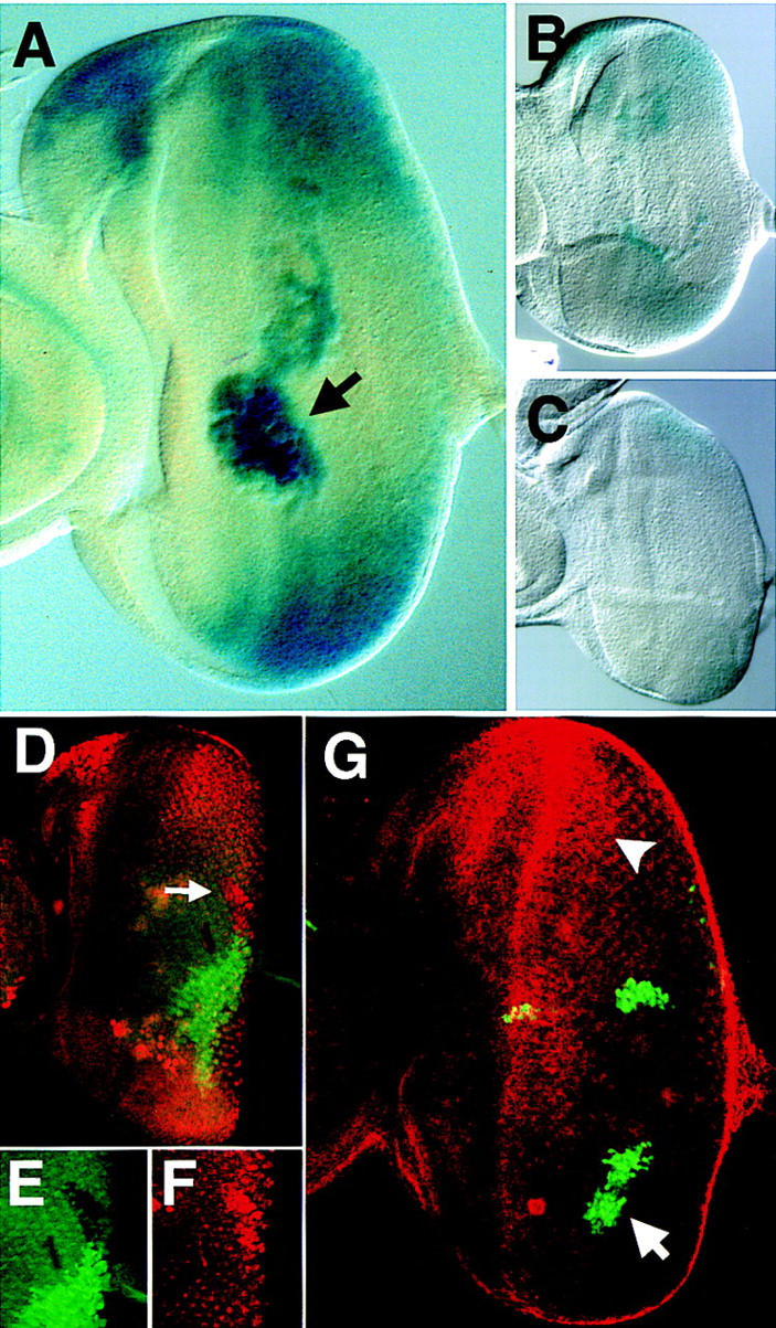
The expression of stat92E–lacZ is negatively regulated by the activity of JAK/STAT signaling. (A) stat92E–lacZ staining in a third instar disc showing highest levels of lacZ at the poles of the eye field, at the ocellar spot, and in macrophage cells located in the middle of the eye disc below the plane of focus (arrow). (B,C) Eye discs prepared and stained under identical conditions carrying stat92E–lacZ (B) and stat92E–lacZ in a hopTuml mutant background (C). (D–F) Double label of eye imaginal disc stained for stat92E–lacZ (red) and a GFP clonal marker (green). A hop2 clone marked by lack of green staining (arrow in D, enlarged in E) and results in higher levels of stat92E–lacZ expression autonomously within the clone (D, enlarged in F). (G) Upd and GFP (green) are coexpressed in GOF clones within the eye imaginal disc also stained to show the pattern of stat92E–lacZ (red). An Upd-expressing clone in the ventral portion of the disc (arrow) completely abolishes the stat92E–lacZ gradient in the adjacent tissue such that no staining is seen at the ventral pole. Staining at the dorsal pole remains strong (arrowhead). Large cells staining in the middle of the disc are macrophages.